
0203 811 8175
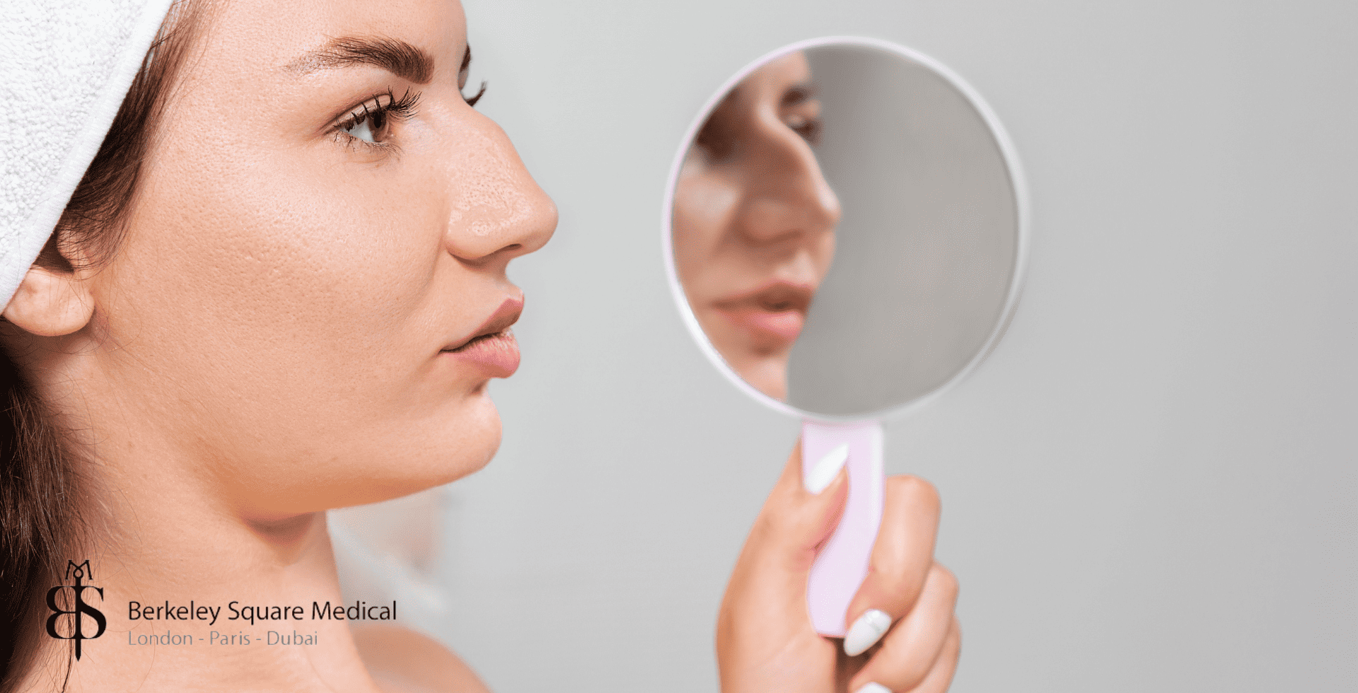
If you’ve had a nose job and, months later, feel like your nose has developed a “beak” shape or a small bump just above the tip, you might be worried about a pollybeak deformity. Patients often describe this as a beak nose after rhinoplasty or feel that the area just above the tip (the supratip) looks too full when viewed from the side.
A true pollybeak deformity is a recognised complication after rhinoplasty, but it’s important to know two things:
In the early weeks, swelling alone can mimic a pollybeak, and this usually improves as healing progresses.
If a genuine deformity develops, it can usually be improved with carefully planned revision rhinoplasty.
This page explains what a pollybeak deformity is, the most common causes after rhinoplasty, and the typical next steps if you’re concerned. It’s for general information only and is not a substitute for a face-to-face assessment with a qualified facial plastic surgeon.
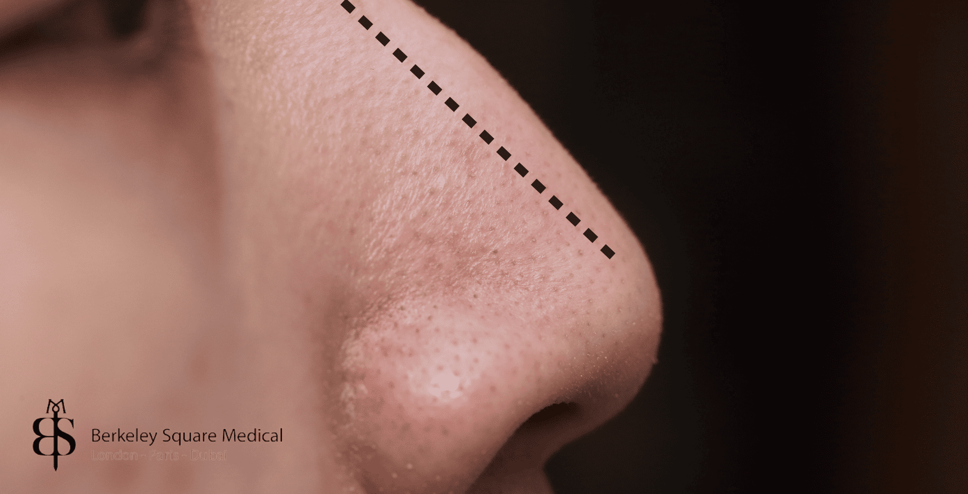
Pollybeak deformity is a postoperative contour problem where the area just above the nasal tip (the supratip) looks fuller than the tip, giving the nose a parrot-beak profile.
It can be caused by under-resection of the dorsal hump, over-resection of supporting cartilage/septum, or excess scar tissue, especially in patients with thicker nasal skin.
Some fullness in the supratip is normal in the first few months after rhinoplasty; a true pollybeak deformity usually only becomes clear 3–6 months or more into healing.
Mild early cases may sometimes be managed with steroid injections or taping, but established deformities typically require revision rhinoplasty by an experienced surgeon.
If you’re worried about a possible pollybeak deformity, the safest step is to discuss it with a specialist rhinoplasty surgeon, who can confirm the diagnosis and explain realistic treatment options.
A pollybeak deformity is a contour problem that affects the supratip area of the nose – the section between the nasal tip and the bridge. Instead of the tip being the most projected point, the supratip looks fuller or more prominent than the tip, creating a profile that resembles a bird’s beak.
Key features include:
Extra fullness or a small bump just above the nasal tip
A tip that looks relatively under-projected or “pushed down”
A profile that appears rounded or “beaky” rather than smoothly sloping
This can happen after both cosmetic and functional rhinoplasty. Sometimes it’s due to the way the nose was reshaped surgically; in other cases, it’s linked to healing, scar tissue, or thick skin. (1)
A pollybeak deformity can have several underlying causes. Often, it’s the result of more than one factor acting together:
If too little cartilage or bone is removed from the bridge during surgery, residual cartilage near the tip can leave the supratip area looking too full. As swelling settles, that extra structure becomes more obvious, creating a pollybeak-style contour.
Sometimes, removing too much from the nasal septum or bridge can cause the tip to drop or lose support over time. When the tip settles but the supratip area remains full, the profile can take on a beak-like shape.
Patients with thick nasal skin are more prone to soft-tissue pollybeak. Even if the underlying cartilage and bone are shaped well, swelling and excess scar tissue can build up in the supratip, masking the surgical refinement underneath.
Every nose heals differently. If the surgeon doesn’t fully account for skin thickness, cartilage strength, or long-term tip support during planning, the final shape may drift towards a pollybeak over several months.
Because the exact cause affects the best treatment approach, proper diagnosis by an experienced rhinoplasty surgeon is essential before considering revision surgery.
It’s completely normal for the nose to look swollen and “full” in the first weeks after rhinoplasty, especially in the supratip area (just above the tip). This early swelling can mimic a pollybeak deformity, even though the final shape has not settled yet.
A rough timeline looks like this:
0–6 weeks: Swelling is at its peak. The bridge and supratip can look puffy or rounded, and it is far too early to judge the final contour.
6 weeks–3 months: Swelling gradually reduces. The basic outline of the nose becomes clearer, but some fullness above the tip is still common.
3–6 months: The nose shape is more stable. If a true pollybeak deformity is developing, it usually becomes clearer during this period.
6–12 months: Subtle refinement continues as deeper tissues settle. Most surgeons prefer to wait close to a year before deciding on any major revision, unless there is an obvious structural issue or severe functional problem.
If you are worried about a possible pollybeak deformity at any stage, the most important step is to raise it with your surgeon at your follow-up visits. They can tell you whether you’re still seeing normal post-operative swelling or whether a genuine supratip contour problem is emerging. In some cases, early interventions such as taping or small steroid injections into the supratip can help manage persistent swelling without surgery.
Treatment depends on what is causing the pollybeak appearance and how long it has been since your original surgery.
If the fullness is mainly due to soft-tissue swelling or scar tissue rather than excess cartilage, your surgeon may suggest:
Supratip steroid injections to soften and thin out scar tissue.
Taping or splinting in the early months to encourage a smoother contour.
Time and observation, as some swelling continues to improve on its own over 6–12 months.
These measures are usually tried first when the underlying bone and cartilage framework is believed to be well shaped.
When the deformity is caused by the nasal structure itself — for example, too much cartilage left near the tip, over-resection of the bridge, or unfavourable scar formation — a revision rhinoplasty procedure is often required. This is typically considered around 12 months or later after the first surgery, once healing has stabilised.
During revision surgery, an experienced facial plastic surgeon may:
Trim or reshape excess cartilage in the supratip area.
Refine the nasal tip framework so the tip projects more clearly than the area above it.
Add cartilage grafts (often taken from the septum, ear, or rib) to rebuild a smooth, natural bridge line if too much tissue was removed previously.
Address scar tissue and re-drape the skin more evenly over the new framework.
Because revision rhinoplasty is more complex than primary surgery, it is essential to choose a surgeon who routinely performs secondary rhinoplasty and has specific experience correcting pollybeak deformity. A detailed consultation, examination, and realistic discussion of goals and limitations will help determine the safest and most effective treatment plan for your situation.
In the first few weeks after rhinoplasty, it’s completely normal for the bridge and tip to look fuller, rounder or slightly “beaky” because of swelling. This can make many patients worry that they already have a pollybeak deformity when, in reality, the nose simply hasn’t settled yet.
A few pointers:
Timing matters – in the first 6–8 weeks, swelling in the supratip area is expected and usually improves steadily.
Texture and feel – swelling tends to feel soft and spongy; true pollybeak related to cartilage or bone often feels firmer.
Trend over time – if the area is gradually shrinking and your profile is improving, it’s probably swelling. If fullness persists or worsens after several months, pollybeak deformity becomes more likely.
Because it’s hard to judge this on your own, it’s essential to attend all follow-up appointments and share your concerns. Your surgeon can examine the internal and external structures, compare to your pre-op photos, and tell you whether you’re seeing normal healing or a developing deformity.
Not every case of pollybeak deformity is avoidable, but the risk can be greatly reduced by careful planning and surgical technique.
Key factors include:
Accurate pre-operative assessment of skin thickness, cartilage strength, and nasal proportions
Avoiding both under-reduction (not removing enough hump) and over-reduction (removing too much support) of the bridge
Ensuring the tip is properly supported and projected, particularly in patients with heavy or thick skin
Respecting the soft-tissue envelope and minimising trauma during surgery
Close follow-up after the operation, with early treatment of problematic scar tissue where appropriate
Choosing a surgeon with a proven track record in rhinoplasty and revision rhinoplasty is one of the most effective ways to lower your risk.
At Berkeley Square Medical, we routinely perform secondary nose surgeries on patients who come to us because they are unhappy with the result of their primary rhinoplasty surgery.
If you suspect that you have pollybeak deformity or are otherwise dissatisfied with the results, book a consultation with us and have your nose examined by our award-winning consultant facial plastic surgeon who is specializing in rhinoplasty.
A pollybeak deformity is a contour issue where the area just above the nasal tip (the supratip) looks fuller than the tip itself, creating a profile that resembles a bird’s beak. It most often becomes noticeable once the majority of post-operative swelling has settled.
In the first few weeks to months after rhinoplasty, supratip fullness is usually due to normal swelling and often improves on its own. A true pollybeak deformity is more likely if the fullness persists beyond 3–6 months, feels firm or structured rather than puffy, and your surgeon confirms it isn’t just residual swelling.
Common causes include leaving too much cartilage or bone in the supratip area, over-resecting the septum so the tip appears relatively over-projected, or excess scar tissue forming under thick nasal skin. Inadequate planning for skin thickness or tip support can also contribute.
Mild supratip fullness caused mainly by swelling or soft scar tissue can sometimes improve with time, taping, or steroid injections. However, structural causes (such as excess cartilage or bone) usually do not resolve on their own and may require revision surgery.
Treatment typically involves revision rhinoplasty. Depending on the cause, your surgeon may reduce excess cartilage or bone, refine and support the nasal tip, or address thick scar tissue. The goal is to restore a smooth, natural bridge and balanced tip profile without over-shortening the nose.
Most surgeons recommend waiting at least 9–12 months after the original rhinoplasty before performing a full revision, to allow swelling to settle and scar tissue to mature. In certain cases of early scar-related fullness, non-surgical options such as steroid injections may be tried sooner, based on your surgeon’s advice.

Patient Journey We are here to guide you to make the right decision for your well-being. If you are considering an aesthetic change to your appearance, we know that it’s one of the most important and personal decisions you wish to make. Therefore you need to be certain that the medical advice you receive is […]

Patient Aftercare All cosmetic surgery procedures at Berkeley Square Medical come with a comprehensive aftercare package and a 24-hour service so you can be assured you are always under the care and guidance of our surgical team. All your immediate aftercare is included in the cost of your surgery so there are no hidden extras in our […]

If you are considering Cosmetic Surgery, we understand that it's probably one of the most important decisions you're likely to make, therefore, you need to be sure it's right for you and you receive professional and independent advice about your procedure.

Our Philosophy We began our journey setting foot in the highly fashionable area of Berkeley Square, Mayfair where luxury fashion, galleries and five-star hotels all conveniently meet to set the example for world-leading client services and experience. Our goal was to create a cosmetic surgery practice that would not only exceed patients’ expectations but provide […]
